
Imaging Anatomy Atlas
Imaging Anatomy Atlas is made and published by 西安欧斯曼科信息科技有限公司. AppBrain is an independent app data site and isn't affiliated with 西安欧斯曼科信息科技有限公司. The app name, icon, screenshots and description belong to 西安欧斯曼科信息科技有限公司 and are shown as published on the App Store.
AppBrain's verdict
- Rating on App Store
- 3.63
- 8 ratings
- Last updated
- September 10, 2023
It doesn't have enough ratings yet to judge what users think.
Rankings
App Store top chartsApp Store Rankings for Imaging Anatomy Atlas
Competitors
Similar apps and more from 西安欧斯曼科信息科技有限公司Alternatives to Imaging Anatomy Atlas






Releases
Updates seen on the App Store- Version
- 1.1.0
- Launched
- July 2023
No changelog data
User feedback
Ratings and reviews on the App StoreComments on Imaging Anatomy Atlas for iOS
We don't have enough comments to show example comments. Please check back later.
Technology
Found in the app by AppBrain- Price
- Free
- App size
- 52.4 MB
- Content rating
- Medium
Technologies used by Imaging Anatomy Atlas
About the app and developer
About Imaging Anatomy Atlas
Imaging Anatomy Atlas is a medical app developed by 西安欧斯曼科信息科技有限公司.
User Ratings
Imaging Anatomy Atlas is rated 3.63 out of 5 stars. The rating is based on 8 ratings.
App Information
Imaging Anatomy Atlas is freely available from the App Store. The app size is 52.39 MB. The latest version available is 1.1.0. The last update was on September 10, 2023.
Technical Requirements
The app has a content rating of Medium Maturity. The app has been available on the App Store since July 2023.
Developer information for 西安欧斯曼科信息科技有限公司
Address:
Store listing
By 西安欧斯曼科信息科技有限公司, from the App Store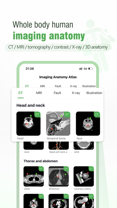
Description by 西安欧斯曼科信息科技有限公司 Written by the developer, not by AppBrain. Click to read.
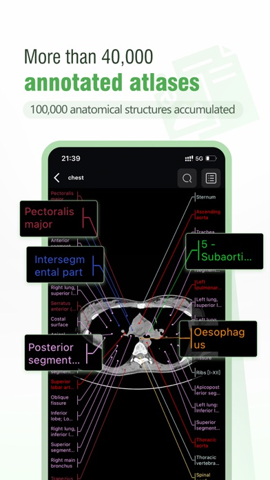
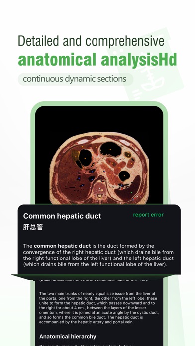
Imaging Anatomy Atlas contains 42,000 medical anatomical images, covering CT, MR, tomography, X-ray, ultrasound, angiography, 3D anatomy and other modules. All images have more than 100,000 anatomical structures to explain. It can learn the knowledge of human anatomy intuitively and comprehensively. Medical imaging knowledge, all kinds of imaging knowledge can be easily obtained.
-- The software has rich and powerful database content:
1. The software has cooperated with major medical colleges and hospitals to collect a large number of 40,000 high-quality imaging maps, covering more than 100,000 anatomical structures, among which a large number of real human anatomy data are high-definition anatomical maps.
2. The classification system is comprehensive, such as upper limbs, lower limbs, spine, head, brain, neck, chest, abdomen, urogenital system image anatomy content, covering X-ray, CT, MR, ultrasound, radionuclide, tomography, 3D anatomy, etc., work and study what you need to learn, super practical, learning images have it is enough.
3. Specific modules include lymph nodes, brachial plexus, Ankle-foot, Coronary artery (angiography), elbow, hand, hip, Lumbar vertebrae, Upper limb (X-ray), Spine (X-ray), Petrous part of temporal bone, Shoulder, Knee (CT arthrography), Lumbar vertebrae, Wrist, Cervical vertebrae (CT arthrography), Face, Male pelvis, Brain, Face and neck, Cerebral arteries, Knee joint, Magnetic resonance cholangiopancreatography, Male human body (tomographic anatomy), Tomography (PET-CT), Pelvic cavity, abdominal pelvis, Head and neck, X-rays, Shoulder (CT arthrography), Female pelvis, Brain, Lower extremity, Shoulder-MRI arthrography, Lower limbs (X-ray), head and neck and extremities CTA, heart coronary VR map, head and neck MRA map; Upper gastrointestinal radiography and urinary IVP radiography.
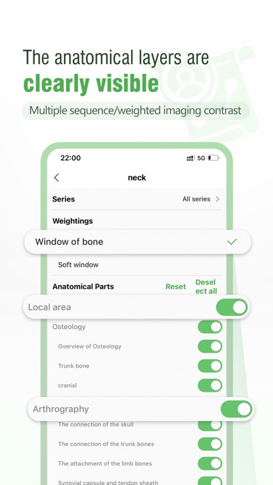
4. Multiple Windows, weighted image differential contrast, software covered T1W, PD-SPIR, T1, T2, Bone, soft tissue, T2W, T2in, Injection contrast media, no injection contrast media, Bone window, Mediastinum, Parenchyma, Computed tomography, Positron emission computed tomography, T1 Gado, Liquid attenuated inversion recovery sequence, DWI and other images.
-- Interactive learning, a new way of image learning
1. The software can toggle the image atlas of each frame by sliding gestures, and support the zoom and zoom of the picture.
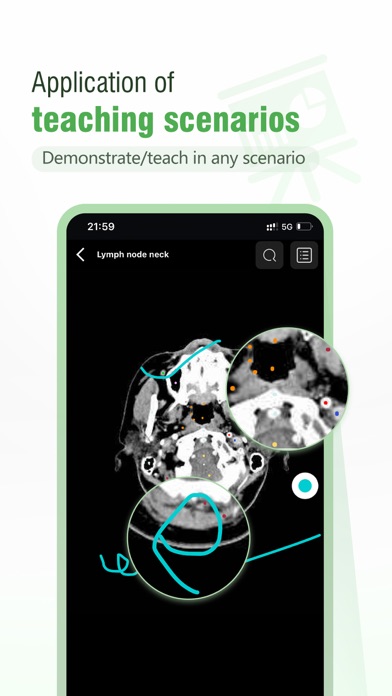
2. The software provides a paintbrush bookmark tool - you can DIY your own notes and support for teaching scenes.
3. Each module can switch different sequences, different viewports, weighted images and filter anatomical structures.
4. You can access the knowledge points of imaging anatomy and read the diagnosis skills and clinical experience of difficult cases at any time and anywhere, learn professional and systematic courses, and quickly improve the diagnostic level and professional skills of primary doctors
Hundreds of thousands of radiologists recommend it. We are working hard to improve and enrich the function and content of the platform. Welcome your use and feedback!
If you have any account/purchase related questions, please contact our customer service at email: anatomyatlas@163.com
We will continue to update the software content